Nikon has just announced the winners of its annual Small World Photomicrography competition, and as you can see from these stunning photographs, bigger isn’t always better.
The competition is in its 42nd year and this year over 2000 people from 70 countries entered. For those that don’t know, photomicrography is the practise of taking a photograph through a microscope or similar magnifying device in order to capture the intricate details of things invisible to the human eye. From the proboscis of a butterfly and the foot of a beetle to espresso coffee crystals, the pictures below give us a whole new way of looking at world. The categories are divided into winners, honorable mentions, and images of distinction, and you can find the full list on the Nikon Small World website.
More info: Nikon Small World (h/t: demilked)
#1 Fourth Place. Butterfly Proboscis

Image source: Jochen Schroeder
#2 Fifth Place. Front Foot (Tarsus) Of A Male Diving Beetle
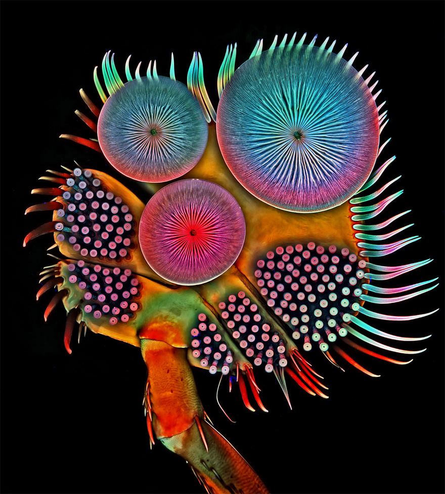
Image source: Dr. Igor Siwanowicz
#3 Eyes Of A Jumping Spider

Image source: Yousef Al Habshi
#4 Nineteenth Place. Human Neural Rosette Primordial Brain Cells

Image source: Dr. Gist F. Croft
#5 Eleventh Place. Scales Of A Butterfly Wing Underside

Image source: Francis Sneyers
#6 Thirteenth Place. Poison Fangs Of A Centipede
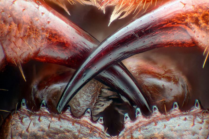
Image source: Walter Piorkowski
#7 Sixth Place. Air Bubbles Formed From Melted Ascorbic Acid (Vitamin C) Crystals
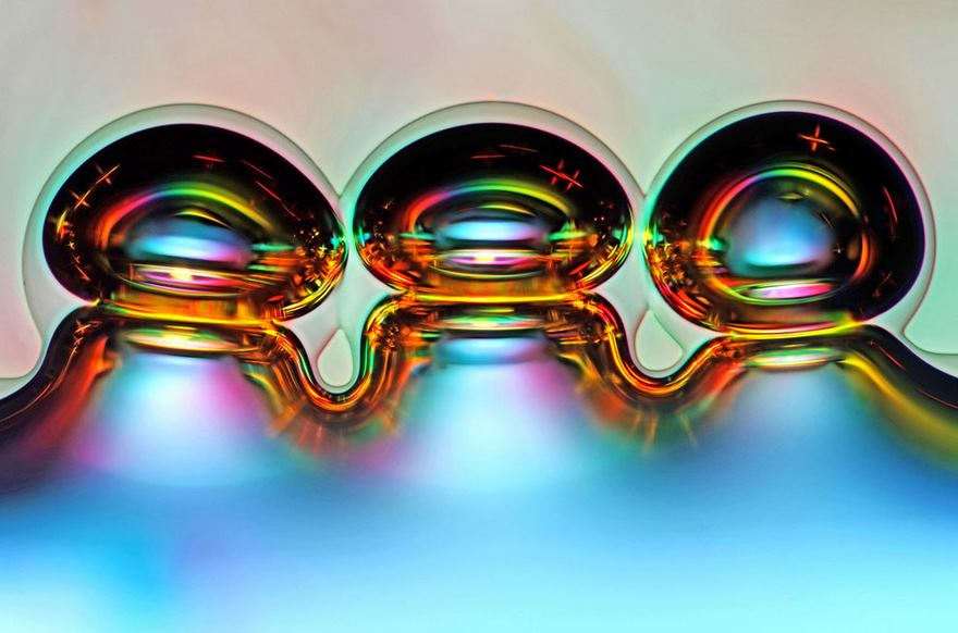
Image source: Marek Mis
#8 Eighth Place. Wildflower Stamens
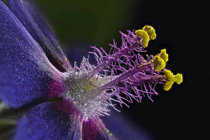
Image source: Samuel Silberman
#9 Retinal Ganglion Cells In The Whole-Mounted Mouse Retina

Image source: Dr. Keunyoung Kim
#10 Ninth Place. Espresso Coffee Crystals
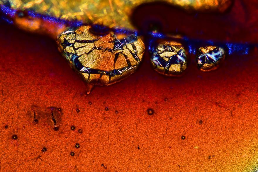
Image source: Vin Kitayama & Sanae Kitayama
#11 First Place. Four-Day-Old Zebrafish Embryo

Image source: Dr. Oscar Ruiz
#12 Caudal Gill Of A Dragonfly Larva
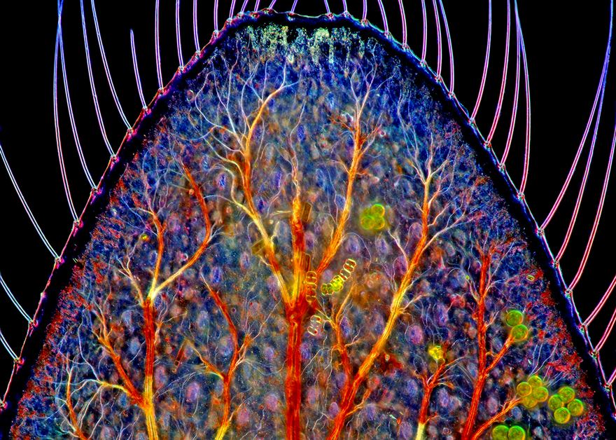
Image source: Marek Miś
#13 Goatsbeard Flower

Image source: Dr. Csaba Pintér
#14 Second Place. Polished Slab Of Teepee Canyon Agate

Image source: Douglas L. Moore
#15 Seventh Place. Leaves Of Selaginella

Image source: Dr. David Maitland
#16 Scales Of A Butterfly Wing

Image source: Evan Darling
#17 Hippocampal Neurons

Image source: Dr. Wutian Wu
#18 Copper Crystals
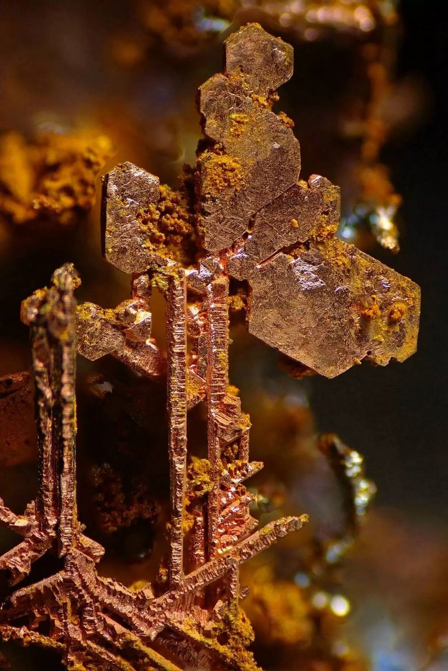
Image source: Honorio Cócera-La Parra
#19 Scales Of A Butterfly Wing
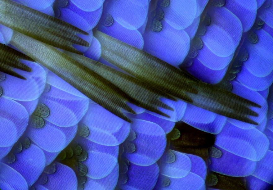
Image source: Anne Gleich
#20 Jellyfish

Image source: Teresa Zgoda
#21 Egg Of A Gulf Fritillary Butterfly

Image source: David Millard
#22 Prolegs Of A Hairy Caterpillar Gripping A Small Branch

Image source: James Dorey
#23 Interference Patterns On A Glycerin Based Soapy Solution

Image source: Haris Antonopoulos
#24 Robber Fly
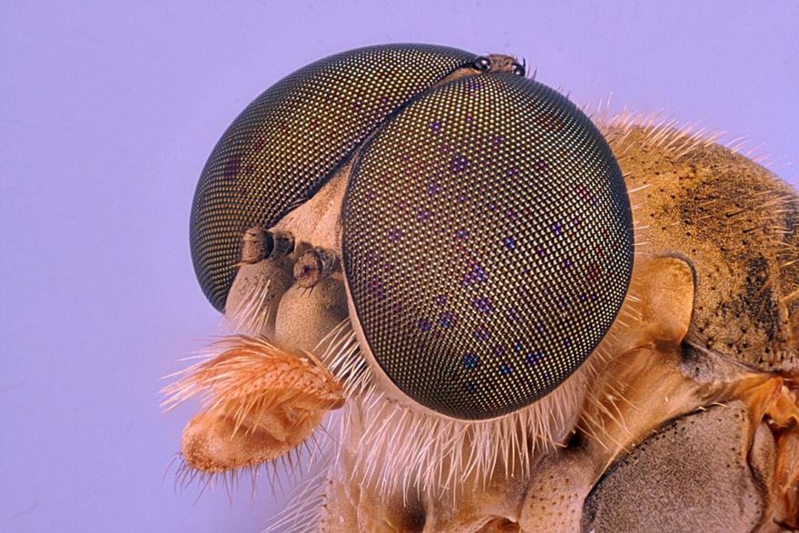
Image source: Jan Rosenboom
#25 Beta-Alanine And Taurine Crystals

Image source: Matt Inman
#26 Gears Coupling Hind Legs Of A Planthopper Nymph

Image source: Dr. Igor Siwanowicz
#27 Twentieth Place. Cow Dung
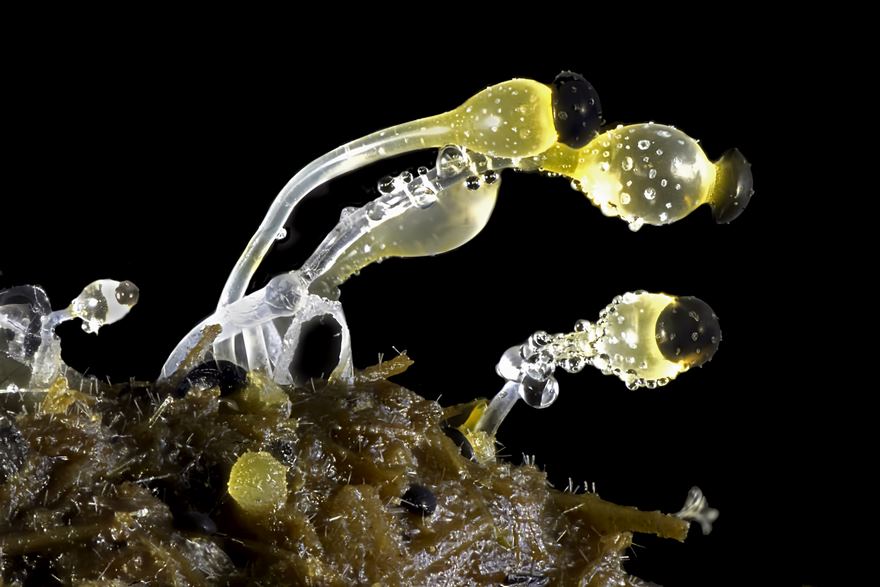
Image source: Michael Crutchley
#28 Water Mite

Image source: Jacek Myslowski
#29 Leg Of A Water Boatman

Image source: Marek Miś
#30 Black Elder Tree Flower Stamen

Image source: Laurie Knight
#31 Ant Leg
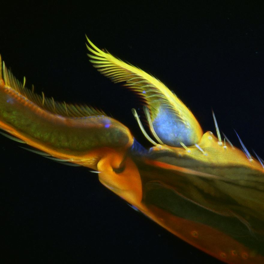
Image source: Györgyi Zséli
#32 Wildflower Stamens
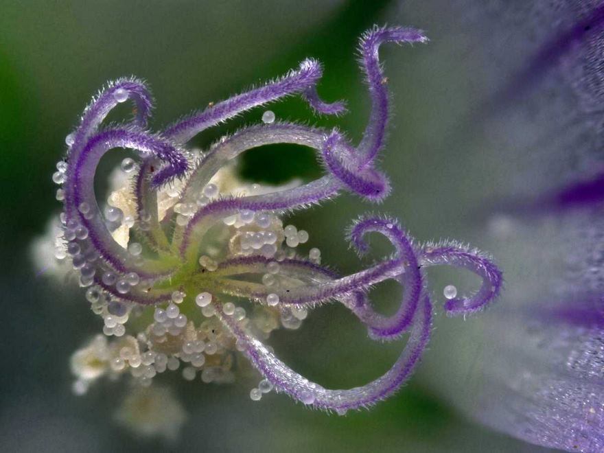
Image source: Samuel Silberman
#33 Twelfth Place. Human Hela Cell Undergoing Cell Division

Image source: Dr. Dylan Burnette
#34 Sixteenth Place. 65 Fossil Radiolarians (Zooplankton) Carefully Arranged By Hand In Victorian Style

Image source: Stefano Barone
#35 Licmophora Flabellata Diatoms
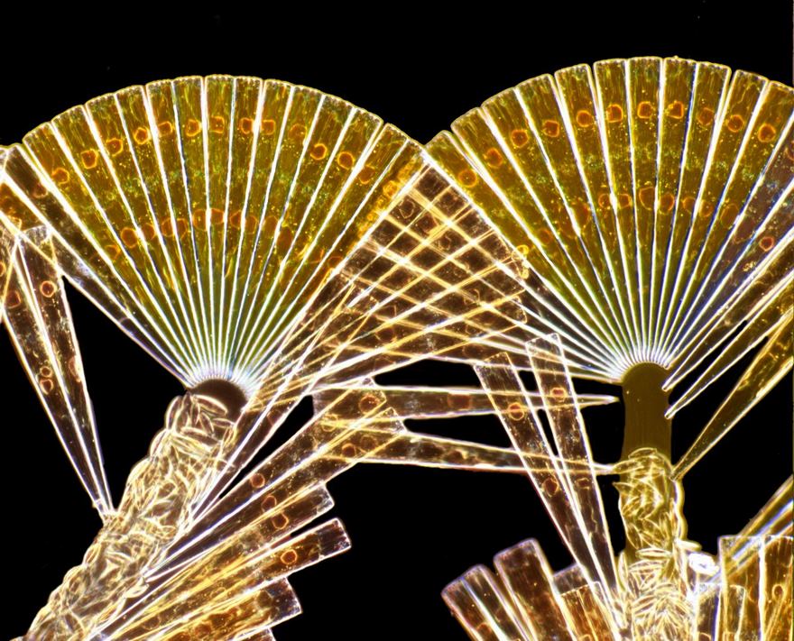
Image source: Dr. Arlene Wechezak
#36 Third Place. Brain Cells From Skin Cells
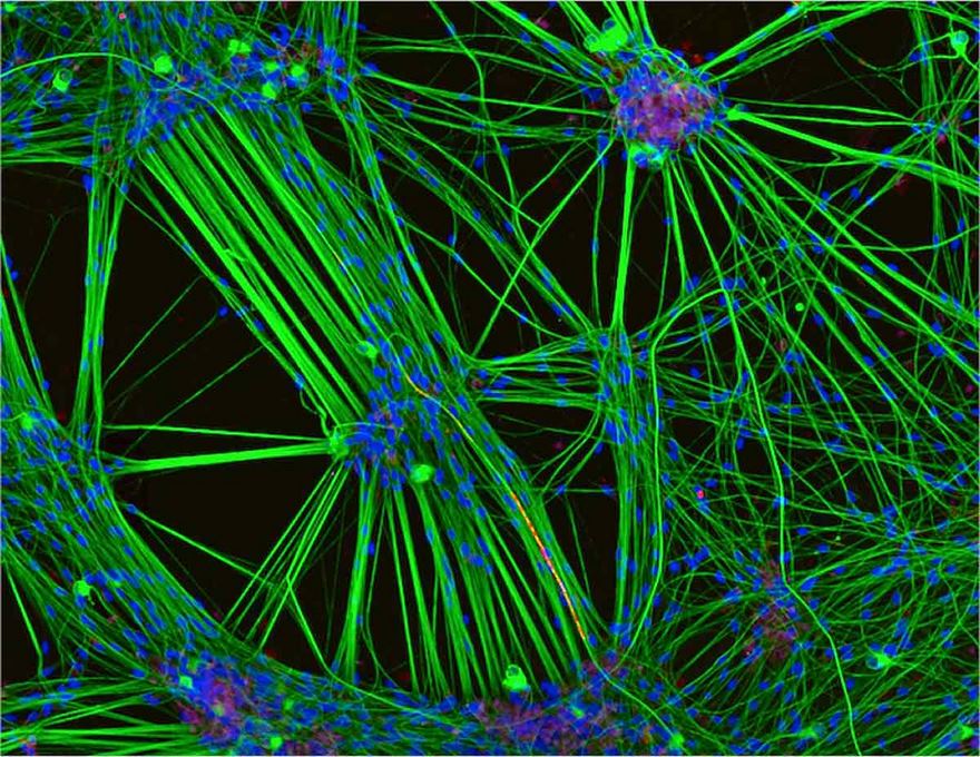
Image source: Rebecca Nutbrown
#37 Spore Capsule Of A Moss

Image source: Henri Koskinen
#38 Green Bottle Fly

Image source: Erno Endre Gergely
#39 Curvepod Fumewort (Corydalis Curvisiliqua) Seed

Image source: David Millard
#40 Seeds Of An Indian Paintbrush Wildflower

Image source: David Millard
#41 Slime Mold

Image source: José R. Almodóvar
#42 Fifteenth Place. Head Section Of An Orange Ladybird

Image source: Geir Drange
#43 Dentate Gyrus Of A Optically-Cleared Transgenic Mouse Brain

Image source: Hei Ming Lai & Dr. Wutian Wu
#44 Testis Of A Fruit Fly
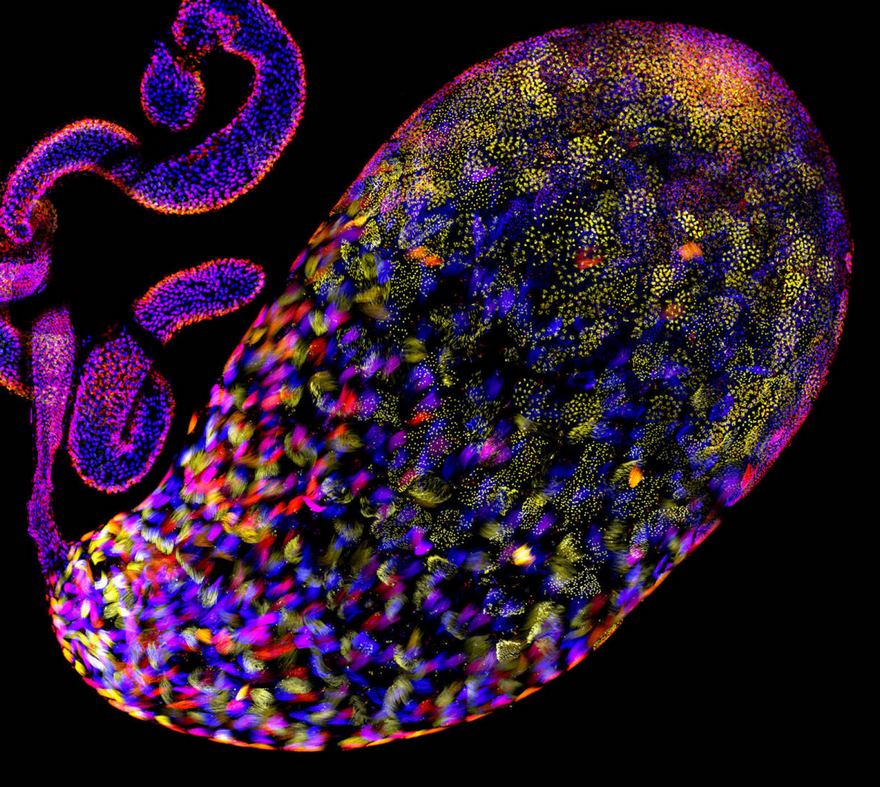
Image source: Christopher Large
#45 Algae
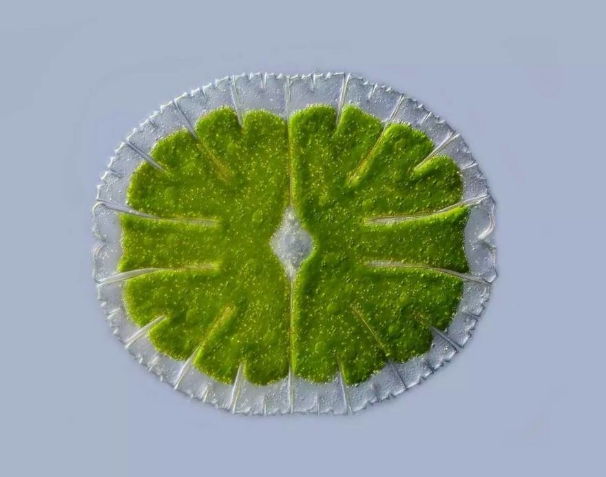
Image source: Anne Gleich
#46 Microcrystal Test For Oxycodone Using Platinic Bromide Solution

Image source: Kelly Brinsko
#47 Ammonite Shell

Image source: Norm Barker
#48 Section Of A Red Speckled Jewel Beetle

Image source: Yousef Al Habshi
#49 Tenth Place. Frontonia (Showing Ingested Food, Cilia, Mouth And Trichocysts)

Image source: Rogelio Moreno Gill
#50 Section Of A Begonia Flower
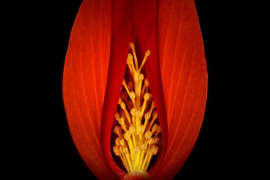
Image source: Viktor Sykora
#51 Tail Of A A Small Shrimp
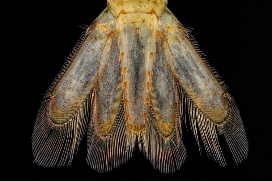
Image source: Charles B. Krebs
#52 Deep Sea Crustacea
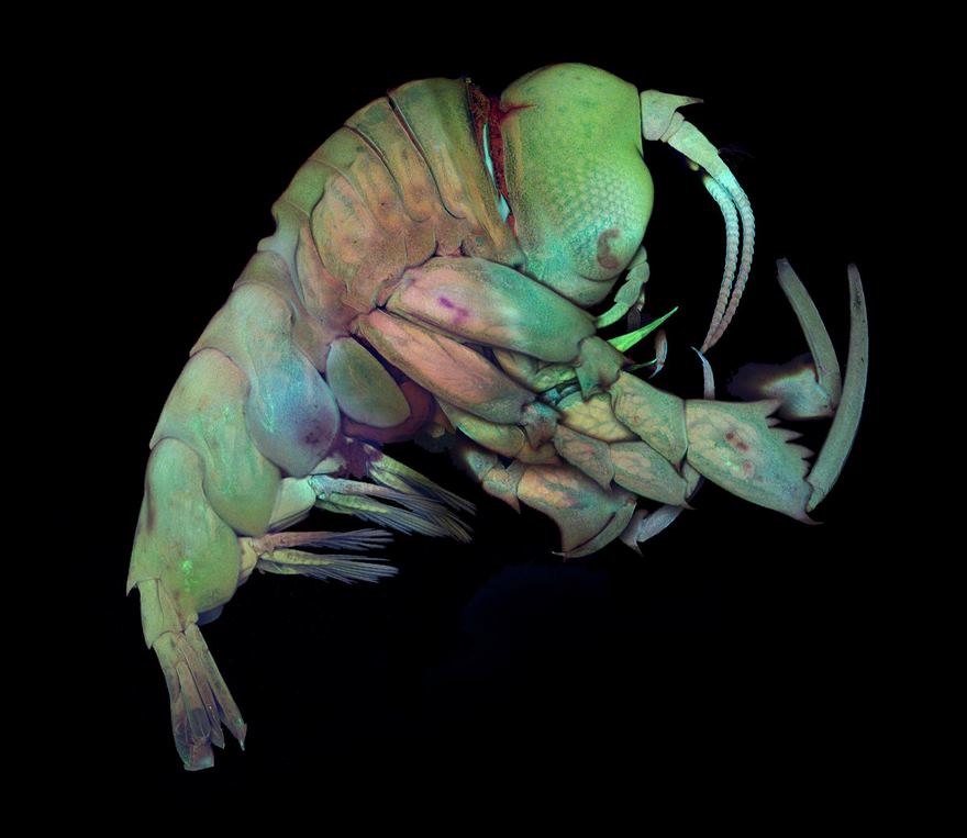
Image source: Dr. Tomonari Kaji
#53 Mouse Hand, Showing Veins

Image source: Evan Darling
#54 Young Flower Buds Of Arabidopsis

Image source: Dr. Nathanaël Prunet
#55 Diclofenac Crystals

Image source: Adolfo Ruiz De Segovia
#56 Ant Pupae

Image source: Geir Drange
#57 Cerebellum Brain Section Of A Rat

Image source: Dr. Barbara Orsolits
#58 Cross Section Of Stem Of Barley

Image source: Dr. Stephen Lowry
#59 Viperfish

Image source: Dr. Alvaro Roura
#60 Early Stages Of Mouse Embryo Development

Image source: Dr. Gaelle Recher
#61 Fourteenth Place. Mouse Retinal Ganglion Cells

Image source: Dr. Keunyoung Kim
#62 Section Of The Cerebellum

Image source: Dr. Marc Leushacke
#63 Galls Of A Mite

Image source: Györgyi Zséli
#64 Surface Of Embryonic Mouse Kidney

Image source: Dr. Nils Lindström
#65 Tintinnid Ciliate Of The Marine Plankton

Image source: Dr. John R. Dolan
#66 Hippocampal Slice Culture Stained For Neurons

Image source: Dr. Jennifer L. Peters
#67 Forewing (Elytron) Of A Tiger Beetle

Image source: Dr. Rudolf Büchi
#68 Leaves Of A Liverwort

Image source: Magdalena Turzańska
#69 Actin, Mitochondria And Dna In A Bovine Pulmonary Artery Endothelial Cell

Image source: Dr. Talley J. Lambert
#70 Mullein Flower
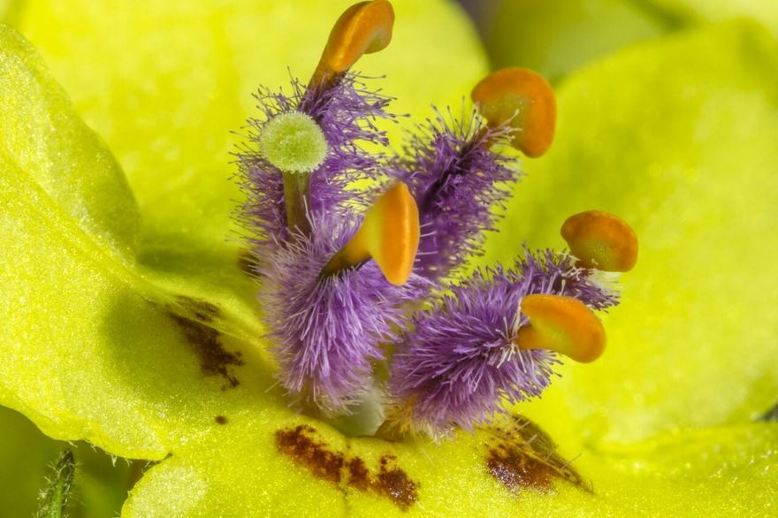
Image source: Karl Gaff
#71 Eighteenth Place. Parts Of Wing-Cover

Image source: Pia Scanlon
#72 Jurinea Mollis Seed
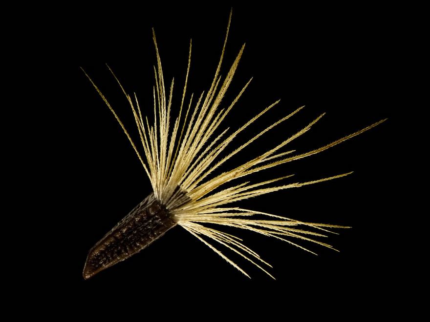
Image source: Viktor Sykora
#73 Micrasterias Thomasiana

Image source: Jacek Myslowski
#74 Moving Vesicles

Image source: Dr. Erdinc Sezgin
#75 Cross Section Of A Lily Of The Valley

Image source: Falco Krüger
#76 Cultured Fat Cells

Image source: Heiko T. Jansen
#77 Cross-Section Through A Multi-Layered Carbon-Fiber Reinforced Composite Structure For Defect Analysis

Image source: Peter Pook
#78 A Daisy’s Central Disc Pattern Of Tiny Unopened Flowers

Image source: Peter Courtney Kinchington
#79 Trumpet Animalcule Containing Endosymbionts

Image source: Wim van Egmond
#80 Dasineura Affinis

Image source: Dr. Csaba Pintér
#81 Mosquito Larva
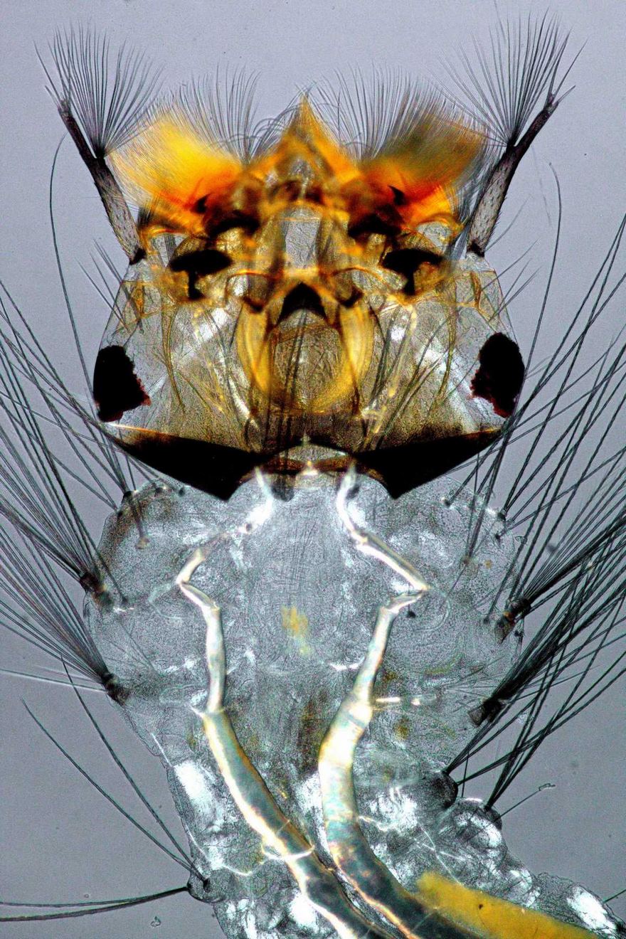
Image source: Edwin Lee
#82 Zebrafish Fin With Cylindrical Bone Segments And Rows Of Pigment

Image source: Dr. Leonardo Andrade
#83 Fossil Diatom From Oamaru

Image source: Frank Fox
#84 Section Of Stem Of A Bomarea Densiflora Plant Specimen
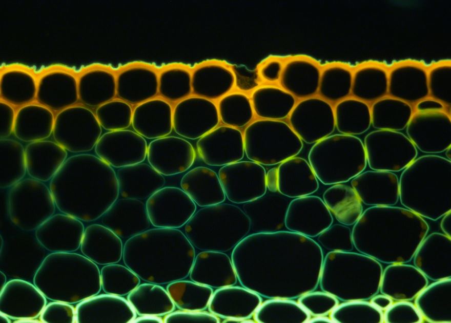
Image source: Edgar Javier Rincón
 Follow Us


