Sometimes bigger is better, but other times it’s all about the details, and as you can see from the winners of Nikon’s 2017 Small World Photomicrography competition, it doesn’t get more detailed than this.
The competition, which is now in its 43rd year, attracts doctors, scientists, and macro photography enthusiasts from all over the world, and over 2000 people from 88 countries submitted their work for consideration this year. For those of you who don’t know, photomicrography is the practice of taking a photograph through a microscope or similar magnifying device in order to capture the intricate details of things invisible to the human eye.
This year’s top prize went to researchers at the Netherlands Cancer Institute, BioImaging Facility & Department of Cell Biology, who captured immortalised human skin cells expressing fluorescently tagged keratin. Scroll down to see the rest of the winners. The categories are divided into winners, honorable mentions, and images of distinction, and you can find the full list on the Nikon Small World website.
More info: Nikon Small World (h/t: mymodernmet)
#1 Jumping Spider, Istanbul, Honorable Mention

Image source: Emre Can Alagöz
#2 Paracetamol (Common Painkiller) Crystals, Finland, Image Of Distinction
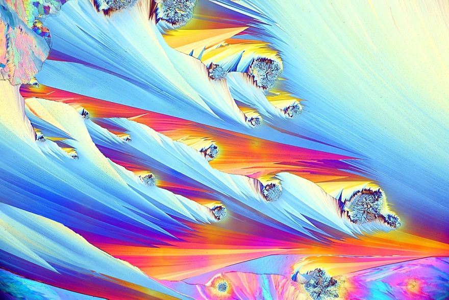
Image source: Henri Koskinen
#3 Living Volvox Algae Releasing Its Daughter Colonies, Nantes, 3rd Place

Image source: Jean-Marc Babalian
#4 The Head Of A Pork Tapeworm, New York, 4th Place
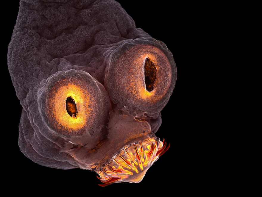
Image source: Teresa Zgoda
#5 Mold On A Tomato, Netanya, 5th Place

Image source: Dean Lerman
#6 Traxacum Officinale (Dandelion) Cross Section Showing Curved Stigma With Pollen, Nottingham, Honorable Mention

Image source: Dr. Robert Markus
#7 Immortalized Human Skin Cells Expressing Fluorescently Tagged Keratin, Amsterdam, 1st Place

Image source: Dr. Bram van den Broek, Andriy Volkov, Dr. Kees Jalink, Dr. Reinhard Windoffer & Dr. Nicole Schwarz
#8 Plastic Fracturing On Credit Card Hologram, Texas, 11th Place
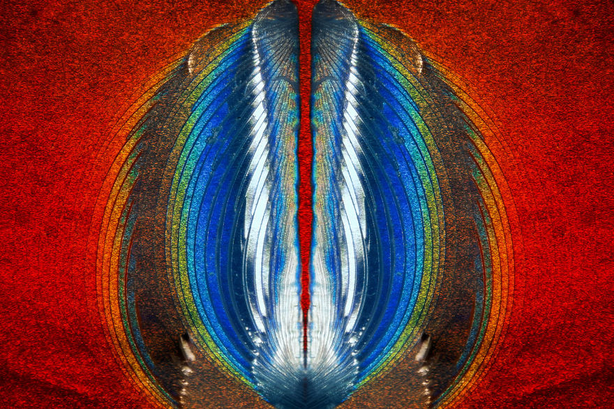
Image source: Steven Simon
#9 Natural Bridge (Petiole Nodes) Connecting The Abdomen And Thorax Of An Ant, Izmir, Image Of Destinction

Image source: Can Tunçer
#10 Synapta (Sea-Cucumber) Skin, Le Mans, 18th Place

Image source: Christian Gautier
#11 3rd Trimester Fetus Of Megachiroptera, Colorado, 15th Place

Image source: Dr. Rick Adams
#12 Asilidae (Rubber Fly) Eye Section, Abu Dhabi, Image Of Distinction
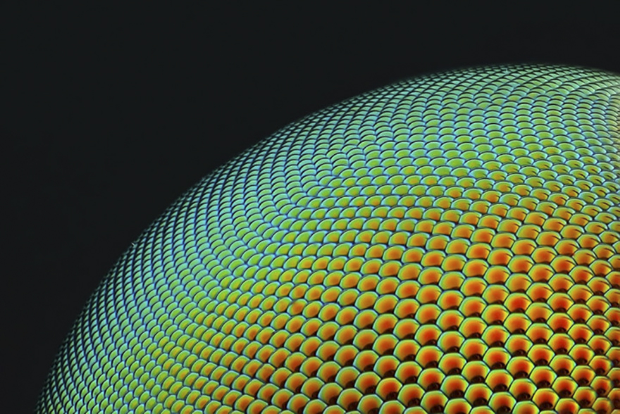
Image source: Yousef Al Habshi
#13 Senecio Vulgaris Seed Head, Israel, 2nd Place

Image source: Dr. Havi Sarfaty
#14 Ganglion Cells Expressing Fluorescent Proteins In A Mouse Retina, California, Honorable Mention

Image source: Dr. Keunyoung Kim
#15 Broccoli, Washington, Honorable Mention

Image source: Dr. Nathan Myhrvold
#16 Opiliones (Daddy Longlegs) Eye, Washington, 12th Place

Image source: Charles B. Krebs
#17 Pyromorphite (Mineral), Spain, Image Of Distinction
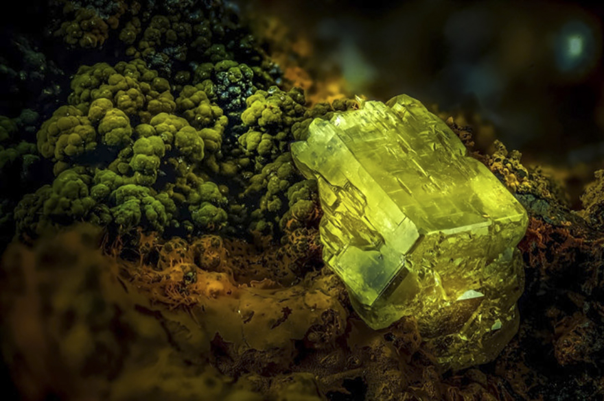
Image source: Emilio Carabajal Márquez
#18 Lily Pollen, Southampton, 6th Place

Image source: Dr. David A. Johnston
#19 Nsutite And Cacoxenite (Minerals), Madrid, Image Of Distinction

Image source: Emilio Carabajal Márquez
#20 Individually Labeled Axons In An Embryonic Chick Ciliary Ganglion, Nagoya, 7th Place
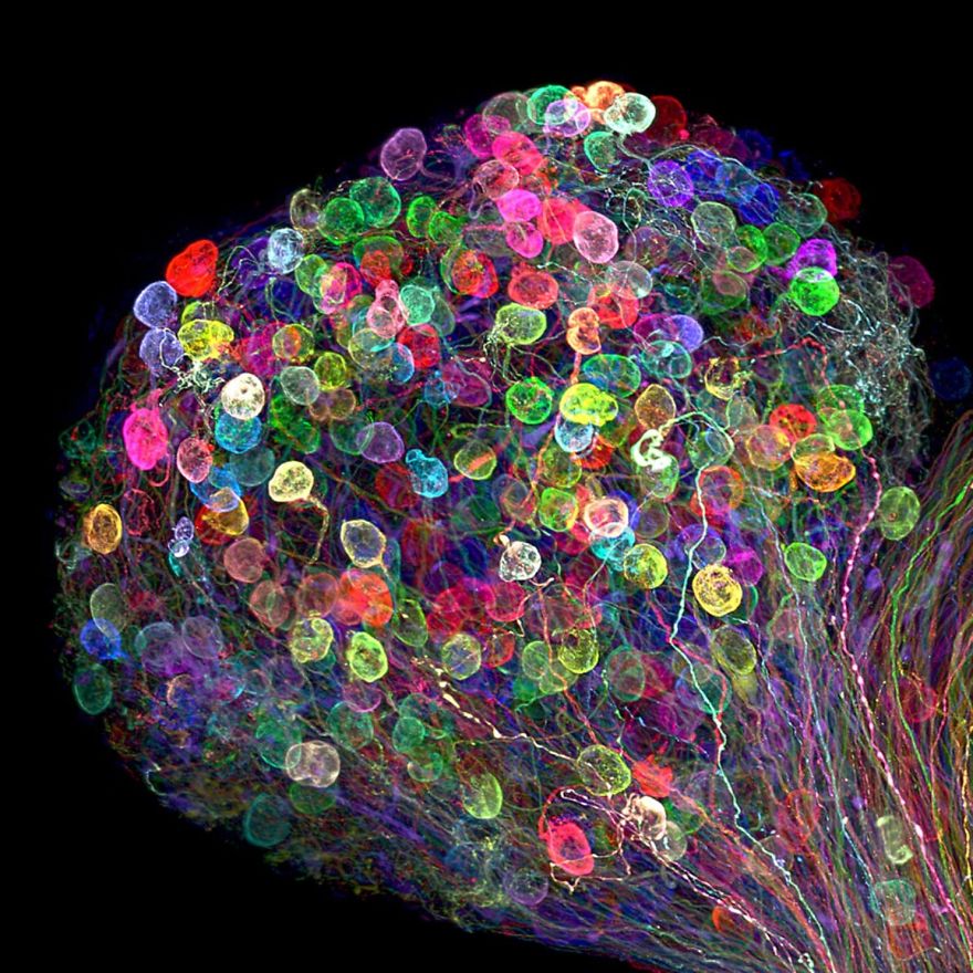
Image source: Dr. Ryo Egawa
#21 Neurons Derived From A Parkinson Patient, Seongnam, Honorable Mention

Image source: Dr. Regis Grailhe, Nasia Antoniou & Dr. Rebecca Matsas
#22 Abdominal Proleg Of A Caterpillar, Netanya, Image Of Destinction

Image source: Dean Lerman
#23 Aspergillus Flavus (Fungus) And Yeast Colony From Soil, New York, 20th Place

Image source: Tracy Scott
#24 Melaleuca Sp. (Paperbark Tree) Leaf, Perth, Image Of Distinction

Image source: Dr. Paul Joseph Rigby
#25 Small Moth, Rostock, Image Of Destinction

Image source: Jan Rosenboom
#26 Moth Eggs In Spider Silk, Illinois, Image Of Destinction
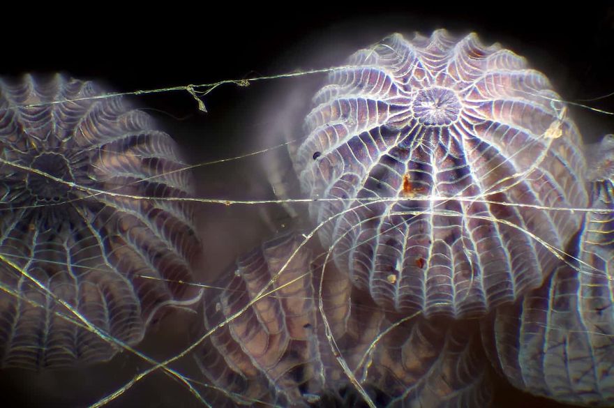
Image source: Walter Piorkowski
#27 Liquid Crystal, Colorado, Honorable Mention
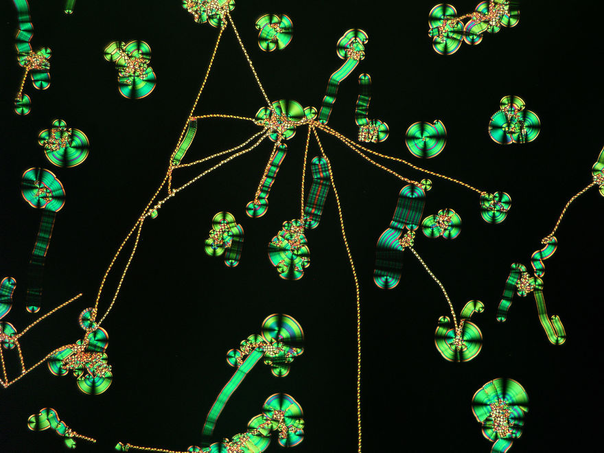
Image source: Michael Tuchband
#28 Parus Major (Titmouse) Down Feather, Podlaskie, 16th Place

Image source: Marek Miś
#29 Nerves (In Green) Under The Skin Of A Mouse (Hair Follicles Are Shown In Red And Blue), Uk, Image Of Distinction
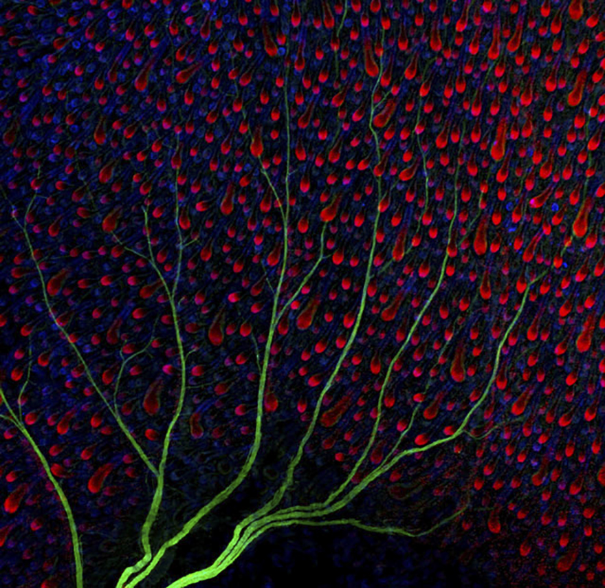
Image source: Henri Koskinen
#30 Dyed Human Hair, Steinberg, 17th Place

Image source: Harald K. Andersen
#31 Natural Sponge, Uk, Image Of Distinction
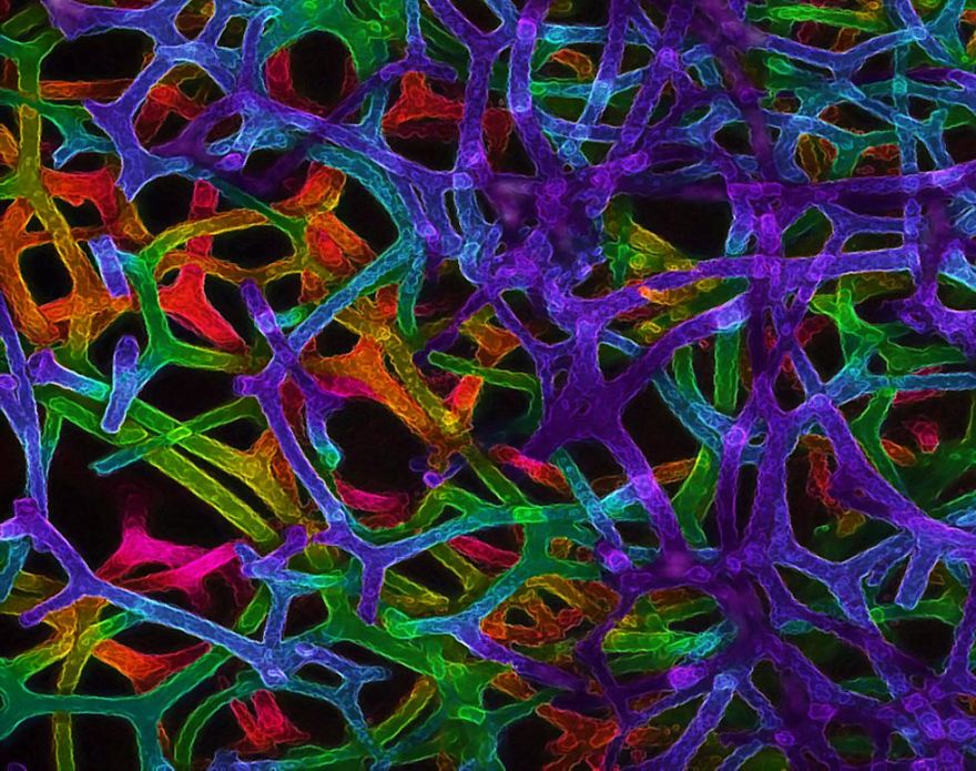
Image source: Dr. David A. Johnston
#32 Common Mestra Butterfly Eggs, Laid On A Leaf Of Tragia Sp., Texas, 14th Place
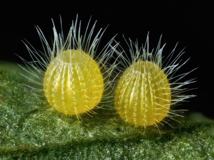
Image source: David Millard
#33 Stamen (Flower Organ),yahud Monsun, Image Of Distinction

Image source: Samuel Silberman
#34 Circocerus (Beetle) Head, Kiel, Image Of Distinction
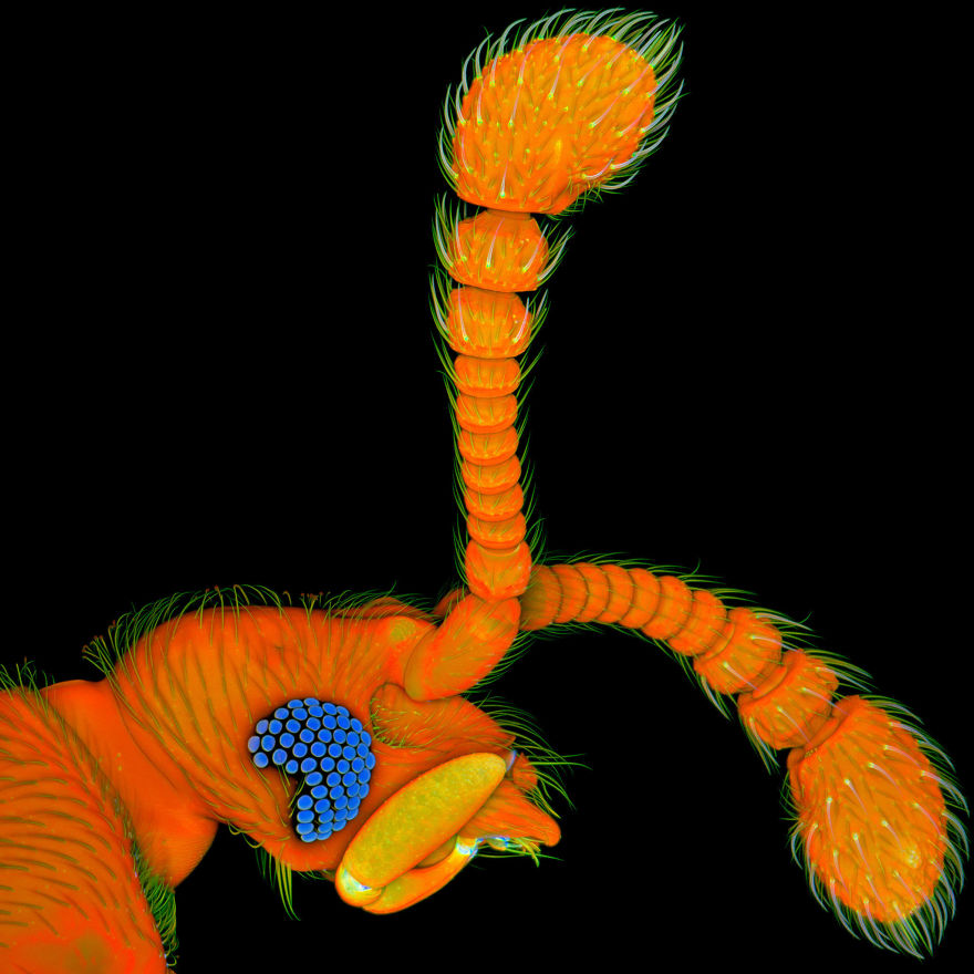
Image source: Dr. Jan Michels
#35 Hippocampal (Brain) Region, California, Image Of Distinction

Image source: Dr. Sarah Moghadam & Dr. Ahmad Salehi
#36 Growing Cartilage-Like Tissue In The Lab Using Bone Stem Cells (Collagen Fibers In Green And Fat Deposits In Red), Southampton, 9th Place
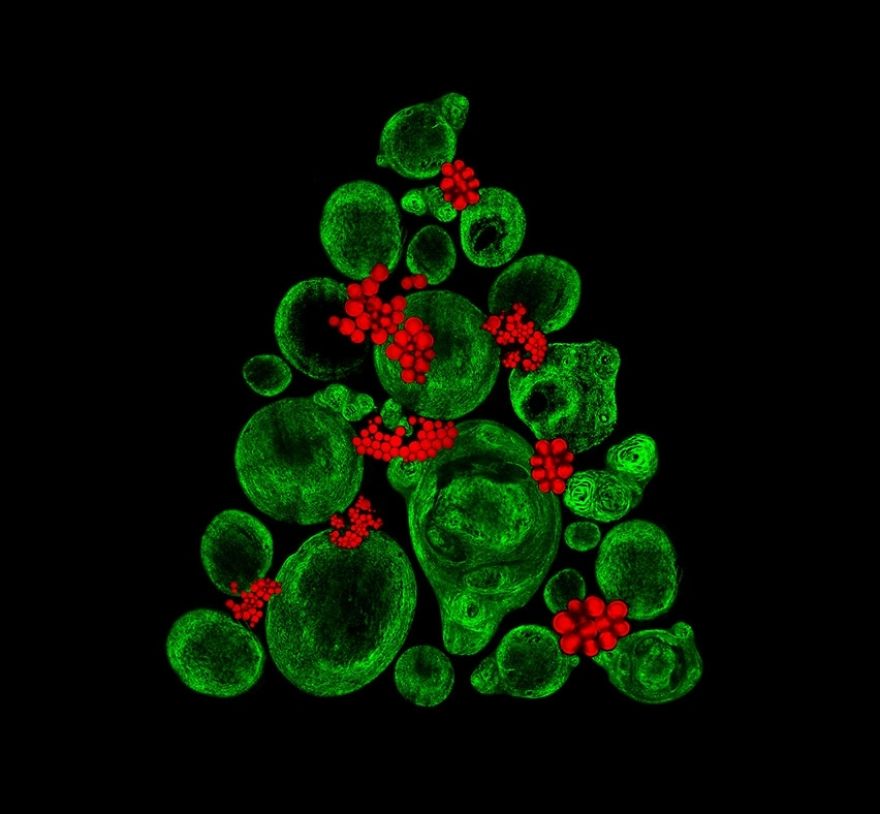
Image source: Catarina Moura, Dr. Sumeet Mahajan, Dr. Richard Oreffo & Dr. Rahul Tare
#37 Newborn Rat Cochlea With Sensory Hair Cells (Green) And Spiral Ganglion Neurons (Red), Bern, 8th Place
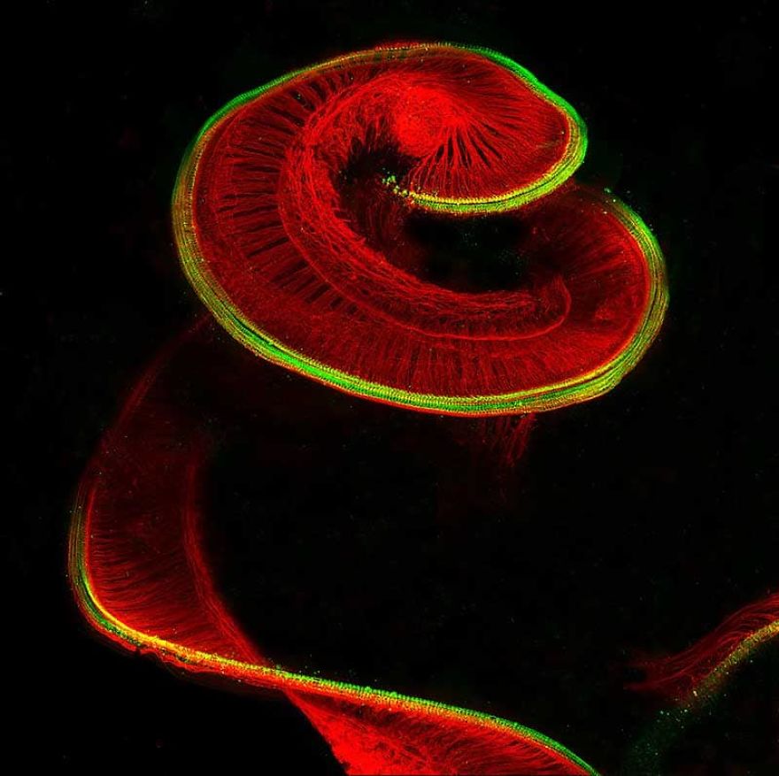
Image source: Dr. Michael Perny
#38 Simple Eyes Of Ectemnius With Condensation, Tonbridge, Image Of Destinction
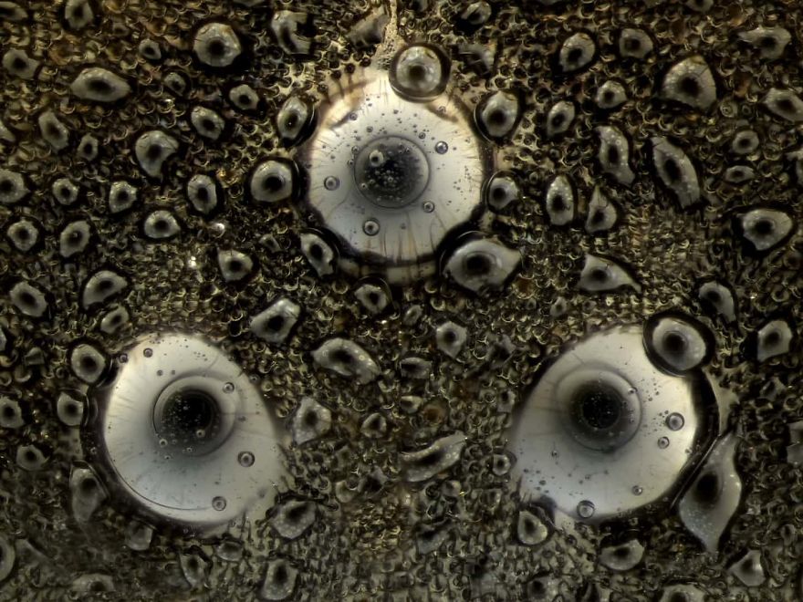
Image source: Laurie Knight
#39 Pleurotaenium Ovatum, Panama, Image Of Distinction

Image source: Rogelio Moreno Gill
#40 Warp Knitted Curtain Fabric, Zwijnaarde, Honorable Mention
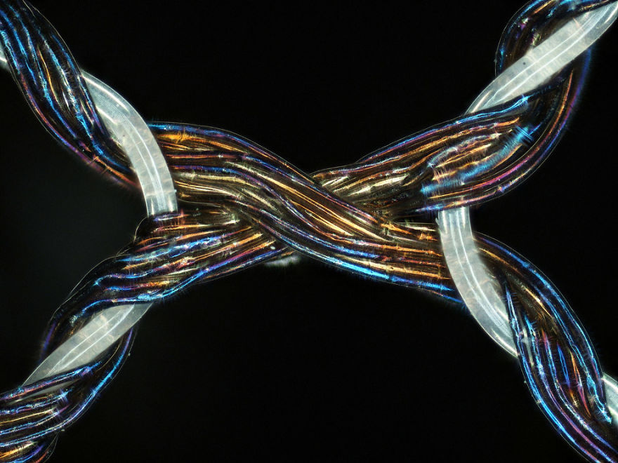
Image source: Marc Van Hove
#41 Label-Free Optical Imaging Of Human Breast Tissue, Illinois, Image Of Distinction

Image source: Sixian You, Dr. Stephen A.J. Boppart & Dr. Haohua Tu
#42 Exaerete Frontalis From The Collections Of The Oxford University Museum Of Natural History, Ramsbury, 13th Place
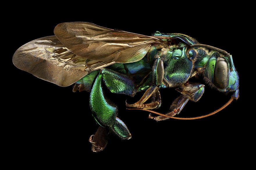
Image source: Levon Biss
#43 Phyllobius Roboretanus (Weevil), Keszthely, 10th Place

Image source: Dr. Csaba Pintér
#44 Migrasome Of A Mouse Fibroblast Cell (L929), Beijing, Image Of Distinction
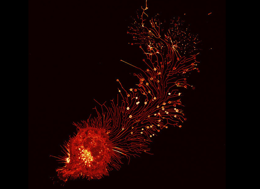
Image source: Dr. Liang Ma & Dr. Li Yu
#45 Human Tongue Blood Vessels Injected With Lead Chromate, New York, Honorable Mention

Image source: Frank Reiser
#46 Sagittal Section Of Mouse Cerebellum (Brain), Athens, Image Of Distinction

Image source: Dr. Aikaterini Segklia
#47 Early Stage Development Of Alcea Rosea (An Ornamental Flower), Usa, Image Of Distinction
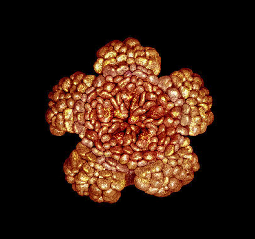
Image source: Masoumeh Sahar Khodaverdi
#48 Prostate Cancer Cells, Usa, Image Of Distinction
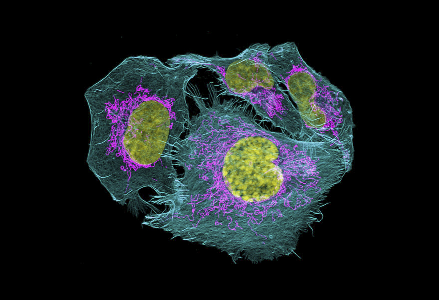
Image source: James E. Hayden, FBCA, RBP
#49 Ciliated Respiratory Epithelial Cells (Yellow) And Mucus Producing Goblet Cells (Cyan), Containing Tight Junctions (Red) And Nuclei (Blue), Rotterdam, Honorable Mention
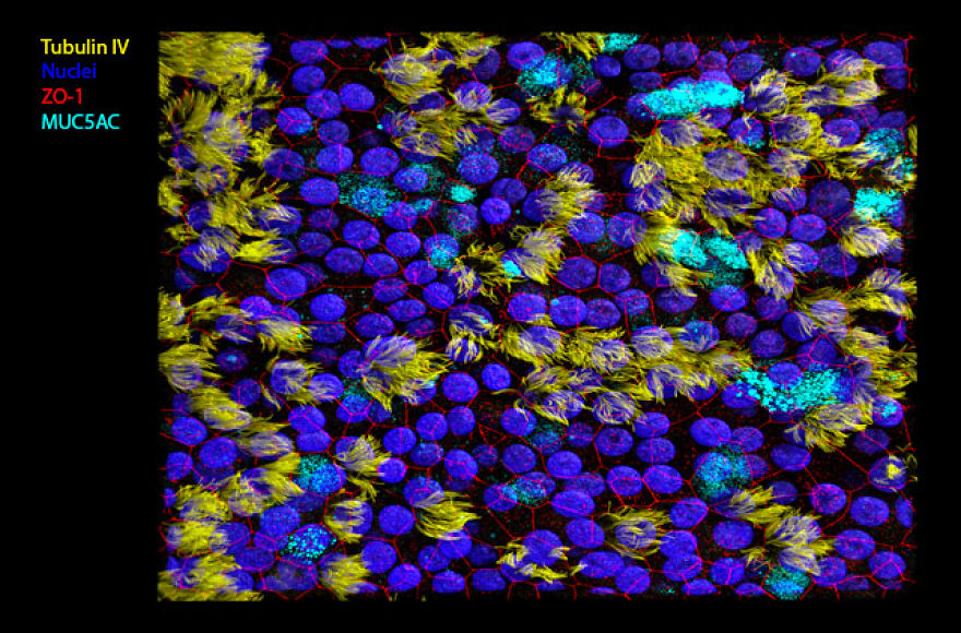
Image source: Alwin de Jong, Dr. G.J. Kremers & Dr. R.L. de Swart
#50 Embryonic Body Wall From A Developing Mus Musculus, Tennessee, 19th Place

Image source: Dr. Dylan Burnette
#51 Dye-Injected Hippocampal Interneuron In Mouse Brain Section, Budapest, Honorable Mention

Image source: Benjamin Barti
 Follow Us


