For more than a century, a tiny structure connected to the female reproductive system was treated as little more than a useless leftover.
The rete ovarii, first described in humans in 1870, eventually disappeared from modern anatomy textbooks. But new research suggests this small structure may actually communicate with the ovary, respond to hormones, and release proteins that could help regulate ovarian function.
The findings have sparked a fresh conversation about how science still has to learn about female anatomy.
“Why has it taken until 2026 for medical researchers to actually FOCUS on female biology/healthcare?” wrote one netizen.
Rete Ovarii has a distinctive horseshoe-like shape and sits around the base of the ovary
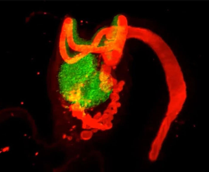
The rete ovarii, often shortened to RO, is a small network of tubes connected to the ovary.
Scientists first described it in humans in 1870. It also exists in several other mammals, including cows, sheep, pigs, dogs, monkeys, and guinea pigs.
For years, however, researchers did not know what it actually did. Because they could not see an obvious purpose, they eventually classified it as vestigial.
In simple terms, that means scientists believed it was a leftover from earlier development that no longer had an important job.
The rete ovarii even appeared in early editions of Gray’s Anatomy, one of the most famous anatomy textbooks. But over time, it disappeared from newer editions and was treated as a functionless part of the adult ovary.
That old assumption is now being questioned.

A research team found that the tiny structure is far more active than expected

In a study published by National Library of Medicine in 2024, scientists studied the rete ovarii in mice to understand its development and possible function.
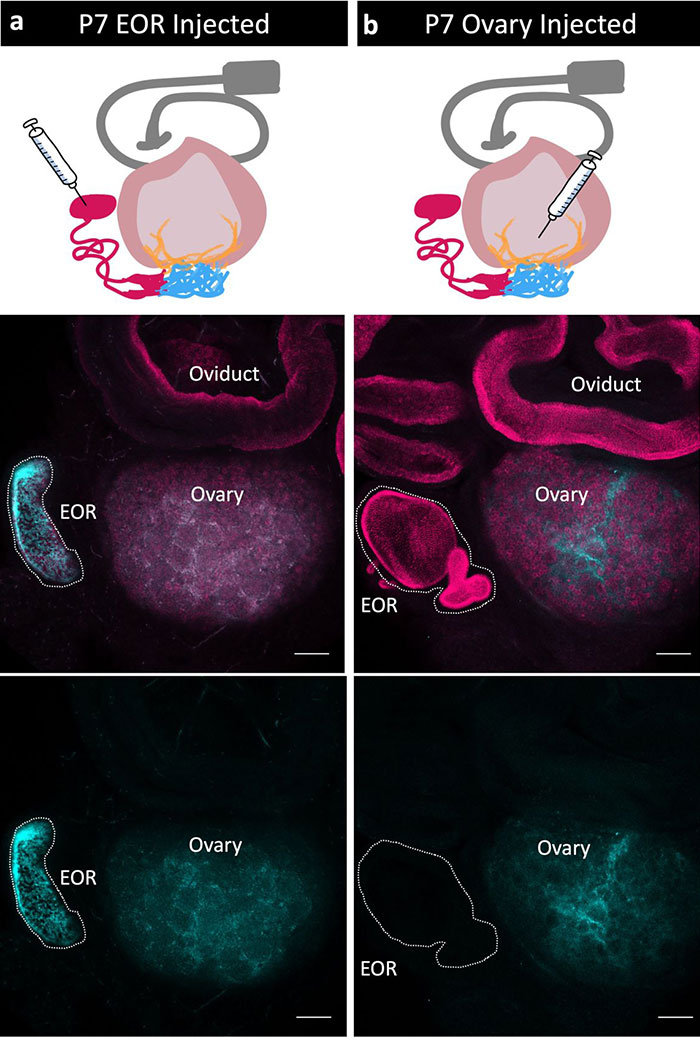
They discovered that it is not one solid piece of tissue. Instead, it has three connected regions: the intraovarian rete, the connecting rete, and the extraovarian rete.
The extraovarian rete, or EOR, is particularly interesting. It forms a twisted tube with a wider end and contains cells that appear able to move material actively.
Researchers injected a fluorescent dye into the EOR to see where fluid moved.
The dye traveled from the rete ovarii toward the ovary. When they experimented in the opposite direction, putting the dye into the ovary, it did not travel back into the rete.
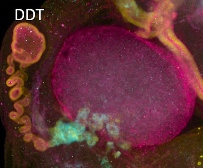
That suggested the structure has a specific direction of fluid movement rather than sitting beside the ovary doing nothing.
The team also found evidence that the rete ovarii produces proteins.
Using laboratory techniques to analyze the material released by the structure, researchers identified several candidate proteins, including IGFBP2. This protein can influence the availability of growth factors involved in ovarian follicle development.
That matters because ovarian follicles contain immature eggs and play an important role in reproduction.
Researchers also found that rete ovarii act like an “antenna” for the ovary
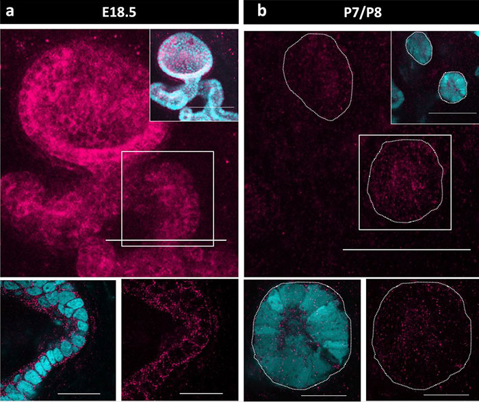


The extraovarian rete sits close to blood vessels, nerves, and immune cells. Its cells also contain receptors for hormones such as estrogen and progesterone.
Those findings led the researchers to suggest that the structure could receive information from different parts of the body and pass signals toward the ovary.
This study described the EOR as potentially acting like an “antenna.”
In simple terms, the idea is that the structure could sense changes involving hormones, nerves, blood flow, or immune activity and then respond by releasing proteins into the ovary.
Researchers have also found that some of the genes active in the rete ovarii change with estrogen levels.
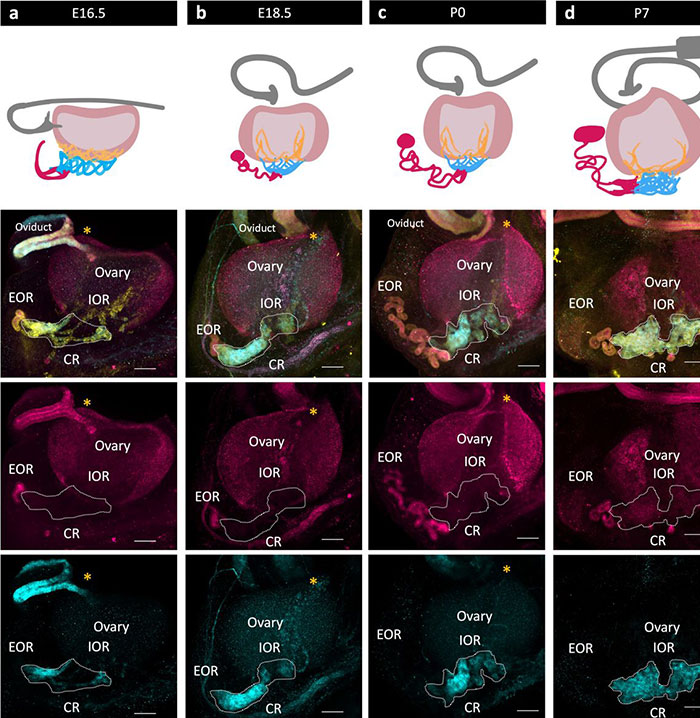
That raises the possibility that the structure responds to the body’s reproductive cycle rather than remaining inactive.
Dilara Anbarci, a developmental biologist at the University of Michigan and one of the researchers involved, said, “There’s still so much we can’t even begin to comprehend about female anatomy.”
She added that she hoped the research would encourage scientists to take another look at what they do not yet understand about the ovary.
The findings are interesting, but researchers have not proved that the rete ovarii is essential for human fertility
The main experiments were conducted in mice, and scientists are still working out exactly what the structure does.
Serge Nef, a developmental biologist at the University of Geneva who was not involved in the research, told Science News that the findings show that “something is going on,” but stressed that it is still the beginning of the story.
Researchers have not yet removed or turned off the rete ovarii in a living mouse to see whether doing so directly changes fertility.
An older study also suggests that normal follicles can still develop when researchers remove the tissue from which the rete ovarii develops early on. That could mean the structure has a supportive role rather than being necessary.
And while humans have been confirmed to have the rete ovarii, scientists do not yet know whether the human structure works exactly like the one observed in mice.
For now, the research shows that the rete ovarii appears to be much more biologically active than scientists once assumed. It does not prove that the structure is the key to human fertility.
The story has also sparked a broader discussion about how medical science has studied female bodies throughout history
Cottonbro Studio/Pexels (not the actual photo)
Adam Taylor, a professor of anatomy at Lancaster University who was not involved in the study, explained that scientists in the late 1800s did not have the technology researchers have today.
“When this structure was first discovered in the late 1800s, it was assumed that it had no purpose because scientists couldn’t see its function within the body,” Taylor told Live Science.
Modern imaging, microscopy and molecular techniques now allow researchers to observe things that would have been difficult to detect in the past, including changes in blood flow and the proteins produced by tiny structures.
That is why scientists can now ask questions about the rete ovarii that earlier researchers could not easily answer.

Kaboom Pics/Pexels (not the actual photo)
The research has also brought renewed attention to other gaps in the history of women’s health research.
The source noted that the National Institutes of Health did not formally require including women as subjects in federally funded clinical research until 1993.
However, the idea that male doctors “deleted” the rete ovarii because they decided it was useless is more complicated than that viral claim suggests.
The historical record showed that scientists struggled to determine the structure’s function, while advances in modern technology have now opened the door to studying it in much greater detail.
“This is the first time I’ve heard of this organ,” wrote one netizen









 Follow Us